Skin temperature is data. Fever raises it, inflammation patterns it, and blood flow writes the signature of vascular health across hands, feet, and limbs. Radiometric thermal imaging reads all of this without contact, without radiation, and in seconds — which is why hospitals use it at the triage desk, and why vascular researchers use it to watch circulation respond to a cold challenge in real time.
This case looks at how one regional hospital deployed the same class of high-sensitivity thermal core in two very different rooms: the outpatient entrance, where it screens hundreds of arrivals a day, and the research lab upstairs, where it is helping map peripheral vascular function.
Project Background
In 2024, a regional hospital in central China — roughly 3,000 outpatient visits a day across its main building — was managing two unrelated frustrations. At the entrance, temperature triage still ran on handheld forehead thermometers: each reading took seconds, required close contact, queued arrivals at peak hours, and produced a number whose accuracy everyone politely ignored. Upstairs, the vascular surgery department’s research group was studying peripheral circulation disorders — Raynaud’s phenomenon, diabetic microvascular changes, early peripheral arterial disease — using assessments that were either subjective (observation, patient history) or slow and appointment-bound (vascular ultrasound). What the group wanted was an objective, dynamic picture of skin perfusion: how fast and how evenly a hand rewarms after a standard cold challenge says a great deal about microvascular function, and thermography is the established way to capture it. The hospital funded a combined procurement: radiometric thermal imaging stations for entrance triage, and a laboratory-grade thermal imaging setup for the vascular research program, built around the same 640×512 high-sensitivity LWIR core.
Pain Points of the Traditional Approach
- Contact thermometry doesn’t scale to a queue. Handheld readings are slow, close-range, and serial; at morning peak the entrance becomes the bottleneck, and accuracy degrades exactly when staff are most rushed.
- A single forehead number is poor data. Skin temperature varies with weather, exertion, and measurement technique; triage decisions deserve better input than one hurried reading.
- Vascular assessment lacked an objective dynamic measure. Observation and history are subjective; ultrasound is static and appointment-bound. Neither captures how perfusion changes over the minutes that matter in a cold-challenge test.
- Research-grade imaging seemed out of budget reach. The lab had assumed quantitative thermography required specialized medical imagers — until the same industrial cores used in precision measurement turned out to meet the sensitivity requirements at a fraction of the cost.
The Thermal Imaging Solution
At the entrance, radiometric thermal stations screen arrivals as they walk past: the system reads facial temperature — anchored to the inner canthus region that correlates best with core temperature — in under a second per person, without contact, flagging only the rare elevated reading for a confirmatory check. Queueing for triage effectively disappeared.
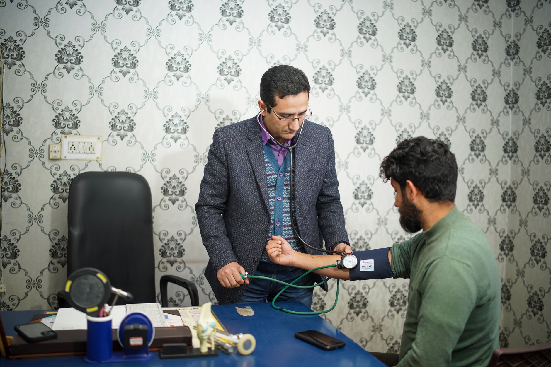
In the research lab, the same class of 640×512 radiometric core, with temperature sensitivity in the tens of millikelvins, images patients’ hands and feet through standardized protocols: baseline mapping, a controlled cold challenge, then a continuous rewarming record. The output is quantitative — rewarming curves, symmetry indices between left and right, region-of-interest temperature tracks — that the group correlates with clinical diagnosis. Because every frame is calibrated radiometric data, results are comparable across visits and across patients, which is what turns interesting images into publishable measurements.
What the Thermal Solution Changed
- Entrance triage stopped being a bottleneck. Screening moved from a queued, contact procedure to a walk-past check; peak-hour congestion at the entrance cleared, and staff returned to clinical duties.
- Elevated readings are confirmed, not acted on. The two-step workflow — thermal flag, then clinical confirmation — cut both missed fevers and false alarms compared with the hurried single forehead reading.
- The vascular group gained its dynamic measure. Cold-challenge rewarming curves now provide an objective, repeatable endpoint for the Raynaud’s and microvascular studies; early results support stratifying patients who previously looked identical on subjective assessment.
- One procurement served two departments. Building both systems around the same core family simplified maintenance, training, and budget approval — a pattern the hospital has since repeated elsewhere.
Module Selection Notes
Medical thermography demands a radiometric LWIR core with high thermal sensitivity — 40 mK class or better — stable calibration, and a digital interface that analysis software can consume frame by frame. The SPECTRA L06A 640×512 LWIR module is the high-sensitivity core for this duty; where a screening station must cover a wide entrance in one view, the higher-resolution SPECTRA L12A 1280×1024 LWIR module fits that role. See the medical application page for screening and research deployment patterns.
Building a thermal screening station or a research imaging setup? Talk to our engineers about core selection, calibration, and software integration.