Research labs have a particular relationship with instruments: the experiment changes every month, the budget doesn’t, and anything bought has to earn its place across projects. That is exactly where an OEM thermal core shines — one radiometric engine that this month maps a charging battery and next month hunts voids inside a brick.
This case covers a university lab that built two very different experiments on the same core-based camera setup.
Project Background
In 2023, the applied-physics lab of a university in Central China consolidated its thermal measurement work onto a single camera built around an OEM radiometric core. The lab serves two very different groups — a battery research team and a civil-engineering NDT team — sharing one instrument budget, and its experiment calendar changes semester by semester. The camera had to be something both groups could script against and carry between benches.
Pain Points of the Traditional Approach
Traditional bench instrumentation forces a trade-off every lab knows well. Contact sensors such as thermocouples give accurate readings at a handful of points, but instrumenting a sample takes hours and still leaves most of the surface unmeasured. Dedicated NDT thermal systems cover the whole surface, yet their cost is hard to justify for a lab whose experiments change every month — and general-purpose cameras rarely export the per-pixel radiometric data that quantitative research needs.
The Thermal Imaging Solution
Experiment 1: Battery Pack Charging Studies
The lab’s battery group studies how cells heat during charge and discharge — where hot spots form, how evenly the pack shares load, and how thermal behavior shifts as cells age. Thermocouples gave them a handful of points per pack; they needed a map.
With the thermal camera recording radiometric video through full charge cycles, the team gets temperature per pixel across the entire pack surface, synchronized with their electrical logging. The outputs are quantitative: time-temperature curves for any region of any cell, exportable as data for their models.
Two practical benefits followed. First, safety: anomalous heating on a suspect cell is visible immediately, which matters when you deliberately push cells to their limits. Second, throughput: a single recording replaces an afternoon of instrumenting a pack with contact sensors.
Experiment 2: Hidden Defects in Building Materials
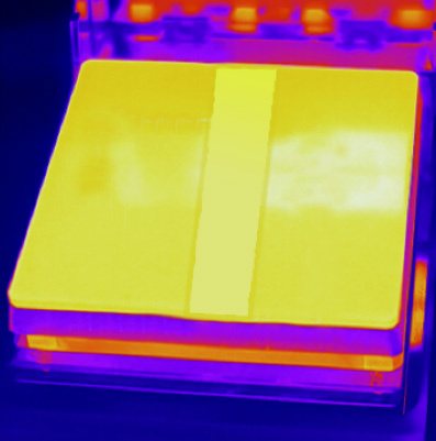
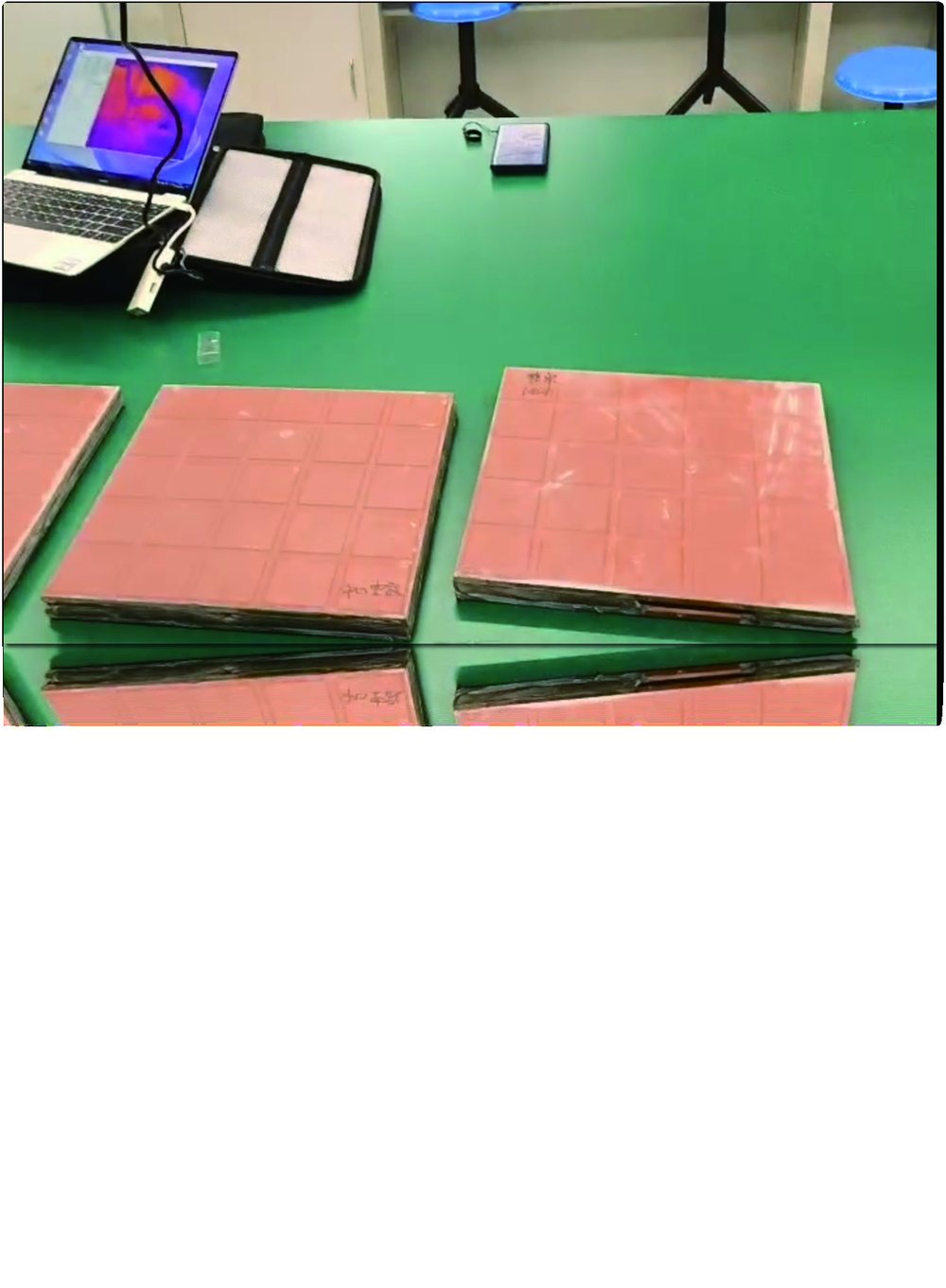
The civil engineering group borrowed the same camera for a different problem: finding cavities and delaminations inside masonry and composite samples. Their method is active thermography — warm the sample surface evenly, then watch how heat flows into it. Sound material warms uniformly; a void interrupts the heat path and shows up as a cool island (or a warm one, depending on the excitation direction).
What used to require a dedicated NDT instrument became an afternoon with a heat lamp, the core-based camera, and their own analysis scripts. The brick samples with known manufactured cavities were located correctly on the first session, which gave the group confidence to move to unknown field samples.
What the Thermal Solution Changed
- Radiometric export. Every pixel carries temperature, so the data feeds directly into analysis scripts and models.
- One instrument, many experiments. Battery mapping this month, NDT next month, electronics thermal validation after that.
- Budget-friendly precision. An uncooled 640×512 core covers most bench-scale work; the lab only outgrows it for very fast transients or very small targets.
Module Selection Notes
For bench research, the SPECTRA L06A 640×512 LWIR core with a close-focus lens covers most material and battery studies; the SPECTRA S06A SWIR core adds a different contrast mechanism for coatings and semiconductor work. More research patterns on the scientific research application page.
Equipping a lab or test bench with thermal imaging? Contact our engineers about cores, lenses, and data interfaces for research use.